Since the eyes are a part of the body, they can be affected by seemingly unrelated health conditions. We must know about your general health and your medications including nonprescription medications prior to examination. A series of special examinations related to the Posterior Segment (Vitreous, Retina, Macula, Optic Nerve, Choroid, Sclera) are available at PrimeonEye for your comfort, namely:
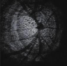
- Swept Source tomography (Latest Technology)
- Fluoro-angiography
- Autofluoresence
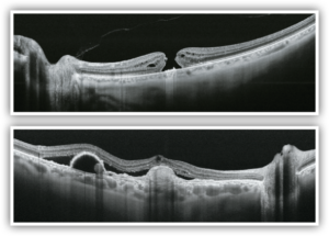
- Macula analysis with 3μm resolution
- Choroid analysis
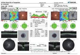
- Glaucoma analysis of Optic nerve, Ganglion cells, Nerve fibers
- OCT angiography of the macula and optic nerve (without dye and without risks) for Macula degeneration, Glaucoma,
- Diabetic maculaopathy, Vein occlusion, etc.
- Fundus true color photography.
SWEPT SOURCE DRI OCT plus TOPCON
Topcon is the first in the world to introduce a combined anterior & posterior Swept Source OCT, the DRI OCT Triton. The DRI OCT Triton incorporates full color high resolution fundus photography and FA & FAF imaging. FA & FAF imaging is a factory option.
Swept Source OCT provides a significant improvement over conventional OCT. Due to the optimized long wavelength scanning light (1,050nm), there is better penetration of the deeper layers of the eye. Furthermore, this scanning light also penetrates better through cataracts, hemorrhages, blood vessels and sclera.
The world’s fastest scanning speed 100,000 A-Scans/second- Approximately twice higher scan speed, compared to Topcon SD OCT, will bring more scans for a single B-scan image, and more informative image supports efficiency and quality of diagnosis.
Better penetration- The high penetration of the Swept Source light can easily and clearly visualize deep layers in the eye, such as choroid and sclera. A further benefit of Swept Source is that it can clearly visualize both the vitreous and choroid in a single scan, that are uniformly clear and noise-free. This eliminates the need for time consuming vitreous/choroidal combination scans.
Wide and deep scans- In one single image the vitreous & choroid are revealed in a crystal clear way. The Topcon DRI OCT Triton enhances visualization of outer retinal structures and deep pathologies. The Topcon DRI OCT Triton automatically detects 7 boundaries including the chorio-scleral interface. The 12mm B-scan covers both the macular area and the optic disc.
Multi modal fundus imaging- The Topcon DRI OCT Triton offers a true color, non mydriatic fundus image while using a very low intensity flash. This unique feature is a perfect tool for identifying the location of scans in the eye utilizing TOPCON’s patented Pinpoint RegistrationTM. The DRI OCT Triton Plus offers a wide range of diagnostic options with multi-modal color fundus imaging, Fluorescein Angiography (FA) and Fundus Autofluorescence (FAF) for even more diagnostic possibilities. For the first time Pinpoint registrationTM will be available with fundus auto fluorescence and Swept Source OCT.

ULTRASOUND b-SCAN
EZ SCAN SONOMED
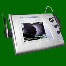
Additional testing may be needed based on the results of the previous tests to confirm or rule out possible problems, to clarify uncertain findings, or to provide a more in-depth assessment. Digital documentation of the front and/or inside of the eyes may be necessary if there are diseases or disorders that need to be reviewed and compared at a later date.
Upon completion of the examination, we will assess and evaluate the results of the testing to determine a diagnosis and develop a treatment plan. We will discuss the nature of any visual or eye health problems found with you and explain available treatment options. In some cases, referral for consultation with another health care provider may be indicated.
Contact us for more advice and help.
